Orbital bone fracture, also known as blowout fractures when the orbital floor has fractured, are in an awkward area in the borders of ophthalmology, ENT, and facial trauma surgery. They may appear as small on the outside and make a big problem on the inside: impincarcerated muscle, incessant double vision, sunken eyes due to being forced to fill an orbital space, vision loss in the worst scenario. The last few years the industry has been evolving silently: imaging, smarter implants, minimally invasive paths, and even machine learning to assist in things that humans sometimes overlook. I will explain the evidence-based updates of recent research (2024-2025) below and translate them into their implications on patients and clinicians.
The fundamentals of orbital fracture
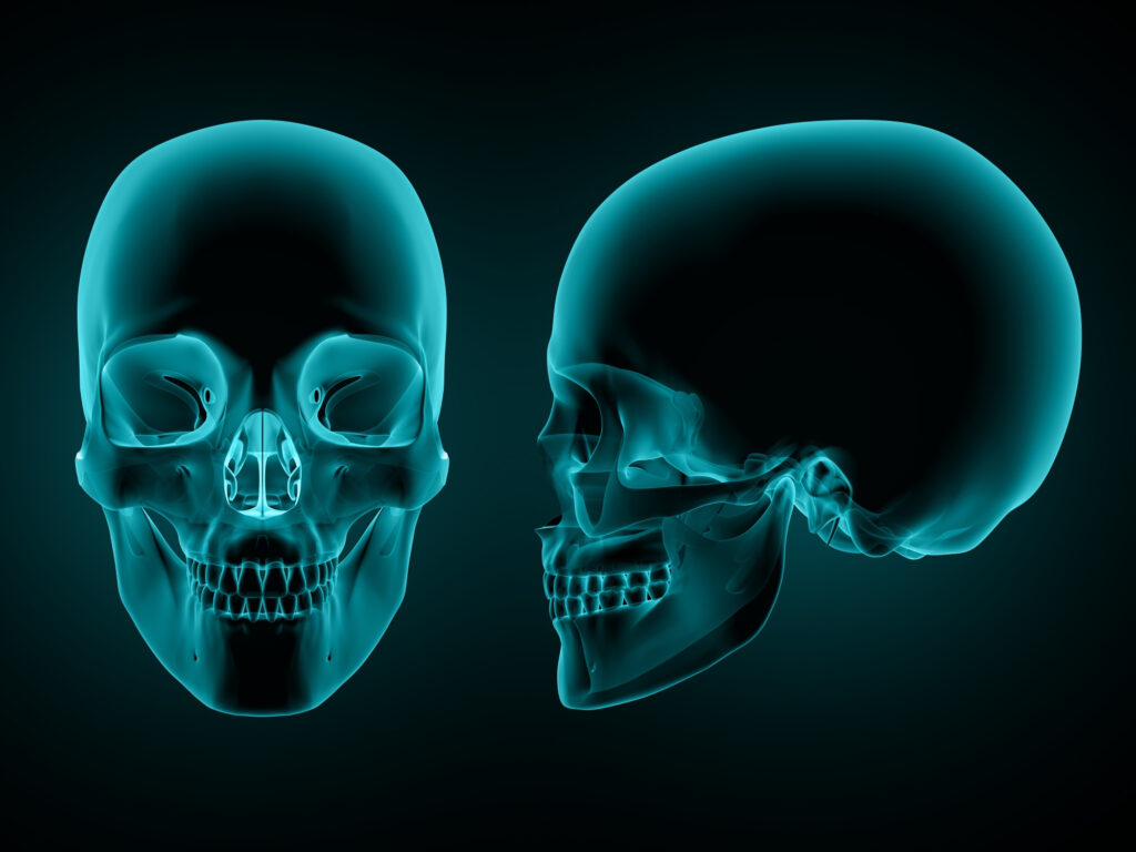
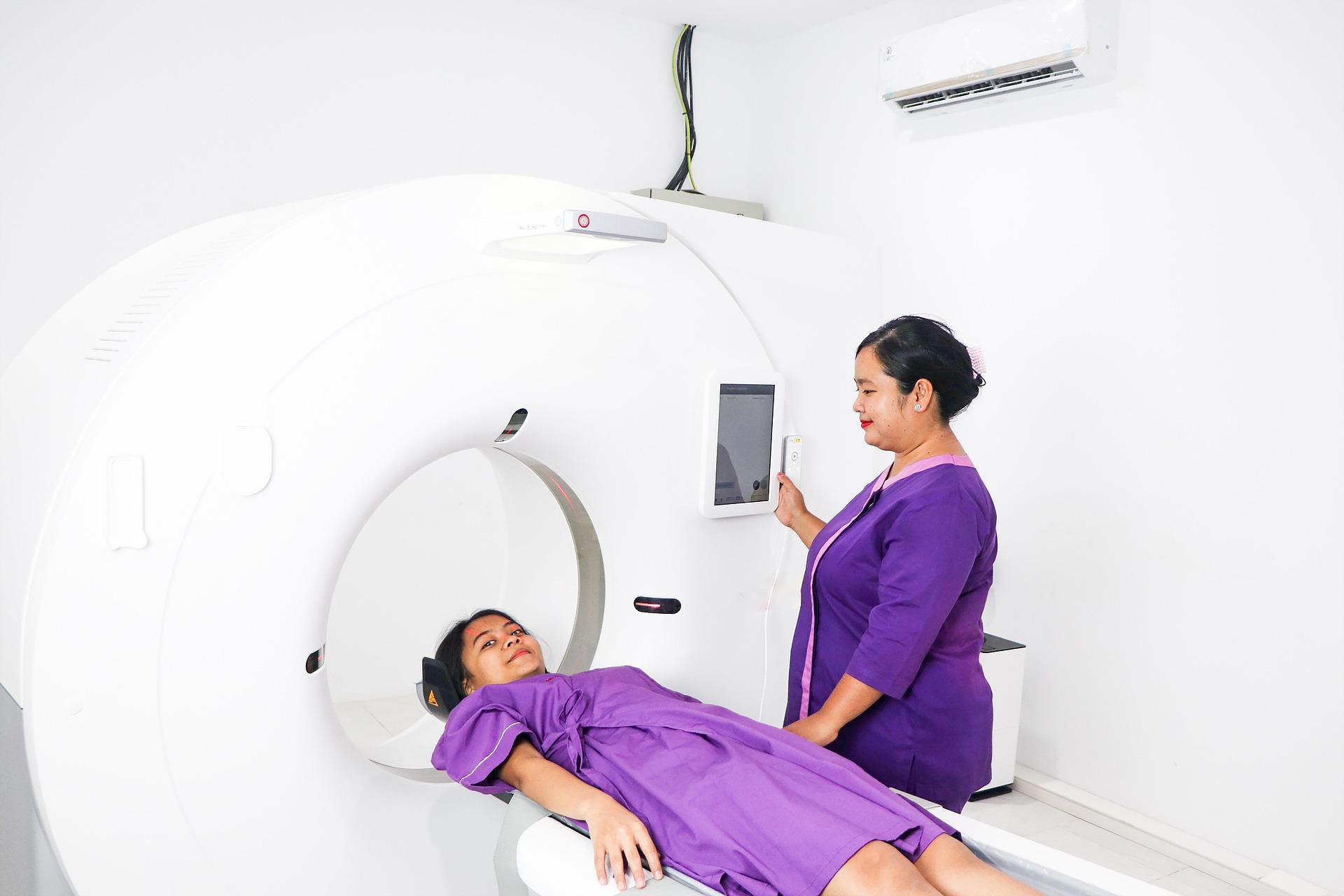
The breaking of bony walls of the eye socket can cause two primary things to damage eye functioning, which include, mechanical entrapment of extraocular muscle or fat within the orbit, and a decrease in orbital volume that may allow the globe to sink (enophthalmos). The symptoms are usually pains, bruising, cheek numbness, restriction in eye movement and diplopia (duplication of vision). The CT scanning is the gold standard of diagnosis as it depicts the bony anatomy and entrapment of soft tissue well. Still, clinical examination and imaging informs what patient should undergo surgery and who may be monitored.
Who requires surgery – and in how short time?
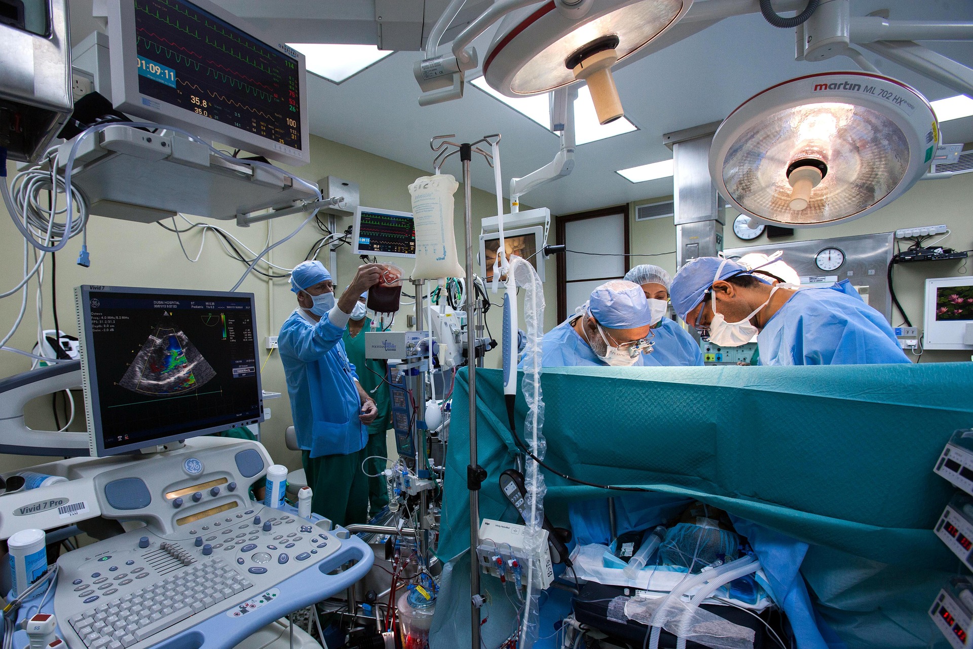
One of the most difficult debates has been timing. Recent reports reinforce a subtle imperative: real emergencies real emergencies, e.g. an orbital trapdoor fracture with muscle trapping, resulting in ischemia; or symptoms of a traumatic optic neuropathy, require exceptionally prompt intervention. In other fractures, multiple period 2024-2025 studies indicate that the range of optimal functional recovery is larger than had been suggested by older dogma, although earlier repair usually benefits those cases with apparent entrapment or progressive diplopia. In 2025, a pooled contemporary clinical experience review suggested that frank muscle entrapment be repaired within 48 hours and large defects that may cause diplopia or severe enophthalmos be repaired within approximately a week; less serious fractures may be observed and repaired at a later stage in case of persistent problems. Shortly: timing is personalized – urgent in case of trapped tissue, timely (in days-weeks) in case of large or functionally important fractures.
Children are not like that, mind the trapdoor
Orbital fractures in children are insidious. The bones of children are more elastic and, therefore, a trapdoor fracture (linear break that snaps together and traps tissue) can happen with minimum external evidence. A series of 2024-2025 pediatrics highlight that despite appearances of subtle CT, features such as nausea, bradycardia or restricted gaze must be causing concern: early surgery in such trapdoor cases results in increased motility and decreased long-term diplopia. Pediatric practice is also more inclined to bioresorbable implants as opposed to adult practice due to the growth factor.

Minimal invasive trends Less cutting, more endoscopes
Endoscopic transnasal and transantral surgeries are no longer an innovative method but a valid instrument in most institutions. The paths enable the surgeons to see the orbital floor either beneath or at the midplane and minimise external incisions and occasionally minimise morbidity. In the recent years, studies demonstrated similar functional results to open techniques in chosen fractures, and new corridors before the lactus majoris and transnasal corridors are increasing access. It does not imply that all orbital fractures must be repaired endoscopically but surgeons have been choosing the path depending on the fracture pattern, presence of other related facial fractures and experience themselves but there are also minimally invasive options that are becoming a tool in the arsenal.
To fabricate your own implants and 3D technology – accuracy reconstruction
Among the most notable changes is the emergence of patient-tailored reconstruction. 3D printing and virtual surgical planning allow teams to build implants that would fit the shapes of the orbital contours of a specific patient. In 2024-2025 clinical series report, orbital volume correction, faster surgeries and better cosmetic and functional results were achieved when patient-specific implants (PSIs) or 3D-printed models used to direct the reconstruction, particularly with complex and delayed cases. Bioabsorbable alternatives (such as constructs based on PCL) are becoming popular especially in the children. The conclusion: it PSI-assisted reconstruction is feasible, it lowers the amount of guesswork and is more likely to increase both the cosmetic and motility endpoints.
You can learn more about Latest Health Updates (2025) Click Here
Imaging and AI – faster, and even smarter, reads
CT will always be central but artificial intelligence will start augmenting the eye of the radiologist. 2025 systematic reviews provide the promise of diagnostic accuracy of deep-learning models that identify orbital fractures in CT, with early systems even able to guide the choice of implant size and shape. These instruments are not yet replacements of clinical judgment, but are similar to a second reader – they are able to identify subtle fractures and more reliably measure changes in orbital volumes than humans alone in certain studies. Anticipate trauma centers to first have workflow integrations, and then general rollouts of models as soon as they are validated. ScienceDirect
Conservative care -in case of observation is suitable
A plate or an implant are not required to all orbital fractures. Non-entrapment small floor defects with limited diplopia not in the primary gaze and no enophthalmos can be treated conservatively: ice, head elevation, short-course steroids in selected inflammatory patients, nasal decongestants, and close follow-up. Current focus is on keen functional hygiene (serial diplopia check, formal motility, and photos to check globe position) as opposed to automatic early surgery. With that said watchful waiting should be not passive but organized – when the problems are not recognized, it is more difficult to delay the repair.
Real-life implications on patients
In case of a blow to the face or a person loved with you or someone else, and the following symptoms appear in double vision, slow movements of the eye, numbing, a cheek, receive immediate clinical assessment and CT. In adults, posing questions to the team would be whether the fracture is entraping mechanically or there is a likelihood of orbital volume change – these two huge surgical flags. When surgically treated, most facilities currently provide minimally invasive surgery and custom-made implants, which are able to accelerate recovery and enhance outcomes. In children, ophthalmology or oculoplastics examination is necessary since trapdoor fractures can require an urgent operation despite mild outward appearances.
Where research is headed
To project into 2025, the following threads will be followed: multicenter studies to determine the most optimal time to perform a non-emergent repair; wider array of clinical validation of AI applications in detection and preoperative planning; randomized trials comparing PSI and non-emerging implants among different fractures patterns; and a longer follow-up period of bioresorbable implants in growing children. Such studies will assist in shifting the expertise opinion to more solid, data-based directions regarding the various types of fractures.
Study more about what is a fractured orbital bone and more by Clicking Here
Sum up bottom line – moderate, contemporary opinion.
The orbital bones fracture are not a unique disease with a specific treatment. The modern technique is customized: the entrapment and optic nerve threats are resolved by a quick reaction, the large or symptomatic defects need a prudent timing, and the implementation of the reconstruction becomes more accurate than ever before thanks to the ever-growing set of tools (endoscopes, 3D planning, patient-specific implants, and AI). To have a simple rule: do not overlook diplopia, or diminished gaze, that is the key to timely, still-saving treatment.
FAQ’s
Just exactly what is an orbital bone fracture and how does it occur?
Orbital bone fracture- One or more bones surrounding the eye socket are broken, often as a result of blunt force – such as in a car crash, athletics injury or not even a fall. Such bones are slim and fragile and a powerful blow may cause them to crack or to give way thus resulting to pain, bruising, and even to have a double vision.
What is the difference between an orbital bone fracture and a tibia bone fracture?
In the orbital bone fracture, the bones that surround the eyes are involved and in the tibia bone fracture, the shinbone- the large bone in the lower leg is involved. The tibia fracture can make one experience a loss of immediate pain and walking ability whereas the orbital fracture only has a significant influence mostly on the vision and eye movement. They both should receive good imaging (CT or X-rays) and occasionally surgery depending on the acuteness.
What are the most typical symptoms of orbital bone fracture?
The commonest symptoms present with a fractured orbital bone are swelling, bruising around the eye, supradiegetic vision, pain on looking in some directions, cheek or upper lip numbness, and in some cases, sunken eye appearance. When you have these following a facial accident, then you need to visit a specialist as soon as possible.
What are the indications of surgery in case of orbital bone fracture?
Orbital bone fracture surgery is typically performed when the victim has muscle entrapment, continuous seeing of two images, or an apparent eye sinking (often referred to as enophthalmos). The surgeon can employ the use of implants or mesh to reconstruct the wall of the eye socket and ensure the eye returns to its proper position. Minor fractures may be effectively treated by rest and observation.
What is the average time spent in the recovery period of the fracture of the orbital bones?
The recovery period of orbital bone fracture is dependent on the intensity of the injury and the presence of a surgical operation. Surgery cases may require 8-12 weeks to recover completely whereas minor fractures require the victim about 4-6 weeks to heal. It usually takes two weeks to go, and then the swelling and bruising will go, but the double vision and the numbness may take more time.
What are the symptoms that a tibia bone fracture requires surgery?
A fracture of the tibia bone may require surgery in case the fragments of the bone are shifted, it is an open fracture (bone protruding through skin), and in case the muscles or nerves are damaged. In 2025, modern facilities are employing materials in surgeries such as titanium nails and plates, which provide quick healing and extend mobility.
Is the orbital bone fracture curable outdoors?
Yes, certain orbital bone fractures recover on their own in case the bones are stable and there is no trapped tissue or significant displacement. Physicians typically follow up scans to evaluate the progress. You probably will be recommended not to blow your nose, make your head stay high, and apply cold compresses to decrease swelling.
What is the painfulness difference between an orbital bone fracture and an injury to the tibia bone?
The pain caused by a fracture of bones of the tibia will be sharper and more painful due to its effects on bone-bearing bones and the fact that it is often fatty tissues, which contain nerves. In has been reported that orbital bone fracture pain is more localized around the eye with most being experienced as pressure or soreness when moving around the eye or even chewing.
Will a surgery on the orbital bone bones impair my sight in the long run?
Normally vision returns to normal following the surgery involving a fracture of orbital bone particularly when this is done early by a skilled surgeon. In unusual cases, minor changes in vision may occur due to scarring or nerve damage, however, with the emergence of 3D-guided approaches and custom implants, which are being developed in 2025, these risks have significantly declined.
What can the physicians do in the case of a orbital bone fracture?
Physical examination and further imaging are conducted together and mostly comprise of CT scan to establish the state of an orbital bone (fracture) injury. They seek any muscle entrapment, bone particles, and an eye position change. Early diagnosis makes the recovery much faster and helps to prevent such complications as having chronic double vision.
Does orbital bone fracture hurt?
Mild to moderate discomfort of post-surgical or post-injury is experienced in most people, and can be treated using medication. The first few days are also characterized by swelling, bruising, and tightness of the eyes that fades away with rest and cold compresses. Taking your aftercare instructions as prescribed by your surgeon will reduce the time you take to get over the orbital bone fracture.
Is it possible to exercise if a tibia or an orbital bone was broken?
In case of tibia bone fracture, light exercises begin when the bone indicates healing and this is usually after a few weeks. Contact sports should be avoided as well as heavy physical activity until the treatment of an orbital bone fracture is monitored by your doctor, preferably in 6-8 weeks so that the injury does not reoccur and that the bones do not experience strain in the healing process.
What would be the consequences of non-therapy of a fractured orbital bone?
The orbital bone fracture may cause a sunken eye or facial asymmetry or permanent double vision when the fractures remain untreated. In exceptional situations, there may be chronic issues with eye movement of the trapped muscles. This is the reason why even the mild cases ought to be examined by an ophthalmologist or maxillofacial surgeon early.
What is the contemporary (2025) method of surgery of orbital bones?
In the year 2025, the majority of the surgeries performed on the orbital bones are minimally invasive. With the help of endoscopic instruments via small holes or via the internal part of the nasal canal, surgeons implant custom-made 3D-printed implants. The procedures decrease scarring, reduce the length of stay in a hospital and enhance cosmetic results.
What would be the contemporary developments in treatment of tibia bone fracture?
The greatest advancements in the management of tibia bone fracture have been smart fixation equipment that enhances quick healing and novel biologic substances that aids in healing. Even AI-assisted imaging is used by some hospitals to predict the course of healing and decide on possible early complications.
Bonus Tip:
In case you have the symptoms of orbital bone fracture, even when it does not appear to be severe, seek medical consultation in the next 24 hours. The fractures located in the eye sockets may appear non-injurious, yet lead to the damage in the long term.